Ct Scan Images Of Head And Neck
The process of taking a ct of the neck begins by taking many different x ray views at various different angles which are then combined with the use of computer processing to create cross sectional images of the bones and soft tissue inside of your body including tissues inside of solid organ. Performed by a radiology specialist this technique can give healthcare professionals an inside look into the neck.
 Ct Scan Of Head And Neck
ct scan images of head and neck
ct scan images of head and neck is a summary of the best information with HD images sourced from all the most popular websites in the world. You can access all contents by clicking the download button. If want a higher resolution you can find it on Google Images.
Note: Copyright of all images in ct scan images of head and neck content depends on the source site. We hope you do not use it for commercial purposes.
Ct scan of head and neck.

Ct scan images of head and neck. Ct scan of head and neck. Welcome to headneckbrainspine a website intended for those interested in neuroradiology anatomy and learning from neuroradiology cases. The most common place for head and neck cancer to spread to is the lungs.
Your doctor has ordered a ct computed tomography of your neck. Other tests that may be done instead of a head ct scan include. The head and neck area is complex and divided into various anatomical and functional subunits.
Positron emission tomography scan of the head. Cancer of the head and neck is the sixth most frequent cancer worldwide and associated with significant morbidity. Also patients with head and neck cancer especially if they arewere smokers can have a separate lung cancer unrelated to the head and neck cancer.
14 public playlist. What are neck ct scans. Radiological anatomy of the head and neck on a ct in axial coronal and sagittal sections and on a 3d images.
Normal ct of the neck. Case ent jan 19. Like traditional x rays it produces multiple images or pictures of the inside of the body.
To navigate the website click on the images below or on the above menu. What is ct scanning of the head. Mri of the head.
Imaging is performed by cross sectional modalities like computed tomography magnetic resonance imaging ultrasound and positron emission tomography computed tomography usually with. Ct scan of head and neck. I have uploaded it so that it can be used for teaching anatomy etc.
The main purpose of this test is to determine if there is a medical problem in the neck area. Also known as computerized tomography or computed axial tomography a neck ct scan is a. Ct of the neck.
Axial c this is a ct scan with contrast of a normal young adult. Atlas of the anatomy of the head and neck on a ct in axial. Ct is an advanced form of x rays that uses a narrow x ray beam and advanced computer software to create detailed cross sectional images of the body organs.
This is one of the safest ways to study the head and neck. The cross sectional images generated during a ct scan can be reformatted in multiple planes. A neck ct scan is a medical procedure where a special type of picture is taken of the neck region.
Your doctor may order a simple chest x ray or ct scan of the chest to investigate. Computed tomography more commonly known as a ct or cat scan is a diagnostic medical imaging test. A ct scan can reduce or avoid the need for invasive procedures to diagnose problems in the skull.
 Ct Scan Of Head And Neck
 Ct Scan Of Head And Neck Showing Parathyroid Cystic Mass Extending
 Ct Scan Of Coronal Section Of Head And Neck Download Scientific
 Ct Scan Of Head And Neck
 A B C The Head And Neck Ct Scan Nov 07 2014 Showed A Tumor 7 2
 Head And Neck Radiology

Effect Of Reduced Z Axis Scan Coverage On Diagnostic Performance

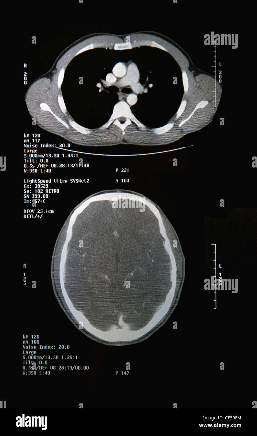 Ct Scan Report Of Head And Neck Close Up Stock Photo 43757832
